|
The
Laboratory |
|
 |
 |
|
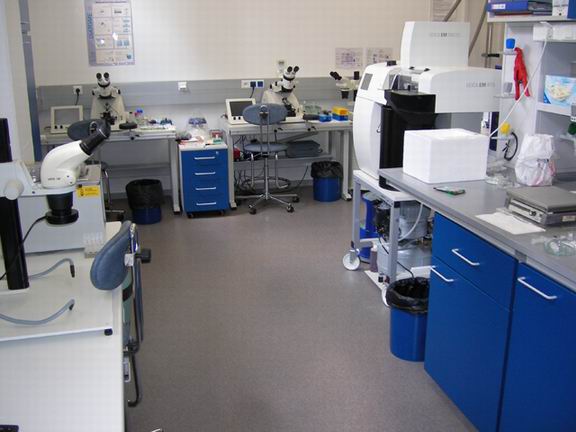
Sample
Preparation for Electron
Microscopy |
|
 LEICA EMPACT2: high-pressure freezing of samples preventing the formation of crystalic ice |
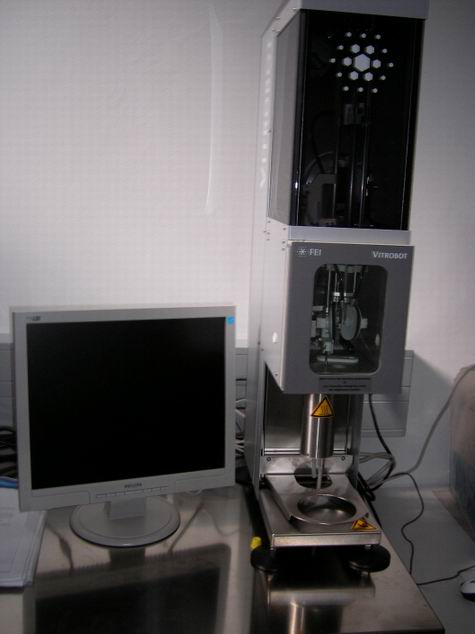 FEI VITROBOT: freezing of samples in thin layers |
 LEICA EM FSP: replacement of ice in the sample with resins |
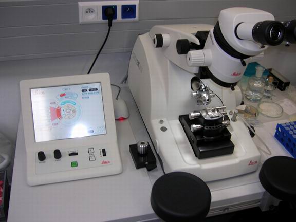 LEICA EM UltraCut6: cutting ultrathin (cryo)sections 50 - 100 nm |
|
Electron Microscopy |
|
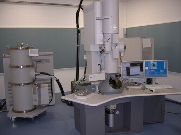 FEI TECNAI: a top research 200kV transmission microscope with tomography, cryostage and elemental mapping (EELS, GIF) |
 FEI MORGAGNI: a routine 100kV transmission EM |
|
Light Microscopy |
|
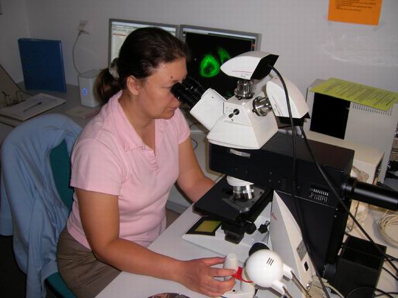 LEICA DM 6000: research grade fluorescence upright microscope |
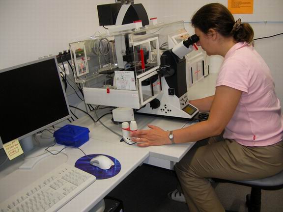 LEICA DMI 6000: research grade fluorescence inverted microscope for observation of living cells |
 NICON TE 300 ECLIPSE: inverted fluorescence microscope for microinjections into living cells |
|