I.
Theoretical background
Light and electron diffusion, optical
systems, waves, reflection, diffraction, interference, polarization.
II.
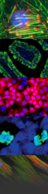
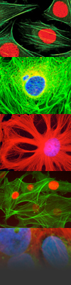
Light microscopy
A microscope and its parts, image
formation, Köhler illumination, optical aberrations, types of lenses,
phase contrast, interference contrast, polarization, fluorescence
microscopy, laser confocal microscopy, two-photon confocal microscopy,
superresolution microscopy, study of dynamic processes in living cells,
immunofluorescence.
III.
Electron microscopy
Characteristics of electrons, resolution
ability, wavelength of an accelerated electron, an electron in a
magnetic field. A scanning electron microscope: design, detection of
secondary and reflected electrons, image creation, X-ray origin and its
use for qualitative and quantitative microanalysis, biological
specimens’ preparation (fixation, dehydration, drying of specimens – a
critical point method, frost preparation methods), SEM image
digitalization. A transmission electron microscope: design, image
creation, interference effects, biological specimens’ preparation
(chemical methods - fixation, dehydration, infiltration, irrigation,
preparation of ultra thin sections, contrasting, physical methods –
low-temperatures processes, microwaves), TEM image digitalization.
Ultra structural immunodetection (immunogold). Comparison of a
photographic and a digital record from a microscope, CCD cameras.
Correlative microscopy. The use of the digitalization and the Internet
in virtual electron microscopy.
IV.
Image processing
1) Image scanning and digitalization
Basic terms (resolution, gray scales,
repeating scanning frequency), advantages and disadvantages of digital
processing, basic ideological scheme of image digitalization. Types of
cameras (analogue versus digital) and their significant features. Types
of capture cards for PC, so called “frame grabbers”, basic principle of
activities. Possible applications and software availability. Concrete
examples of configuration (potential suppliers) and resolving of some
typical problems. Image parameters: contrast, image noise, histogram.
Densitometric calibration. Formats of data files (binary, gray-scale,
RGB, HSV, Lab) and compression (dissipative, non-dissipative).
Filtering and image processing.
2) Basic methods of segmentation
Areas detection: thresholding and areas
growth, edge detection: operators highlighting contours (Sobel, LoG,
DoG), active contours.
3) Measurement of geometric features of a
digital image
Interactive methods: location, length, profiles,
histograms in ROI. Usage of Croft’s formula for measuring circumference
in 2D. Interactive stereologic methods – STESYS system and automatic –
surface, circumference, Feret diameter, quantity, Euler characteristic.
Effect of object anisotropy and noise on measurement accuracy.
4) Image analysis and
visualization in 3D
CLSM and MRI data
resources, dimensional calibration. Filtering and data segmentation.
Usage of Croft’s formula for measuring the surface and length in 3D
measurements. Interactive stereologic methods: Fakir probe and Slicer,
automatic: capacity, quantity, surface and length in 3D. Visualization:
volume and surface rendering.
V.
Stereology and morphometry
Traditional
morphometric methods: measurement of length, surface, circumference,
and quantity. Introduction to stereology. Sampling in stereology.
Cavalieri principle for capacity measurement, dot method. Examples of
stereologic methods for measuring capacity, surface, length and
quantity. Methods for measuring length and surface of 3D structures
from thin sections: a method of vertical sections, orientator. Methods
based on sharpening thicker sections: disector principle for counting
3D particles (e.g. cells), methods for length measurement of space
curves (e.g. capillaries) and surface of 3D structures.

 Course sponsors